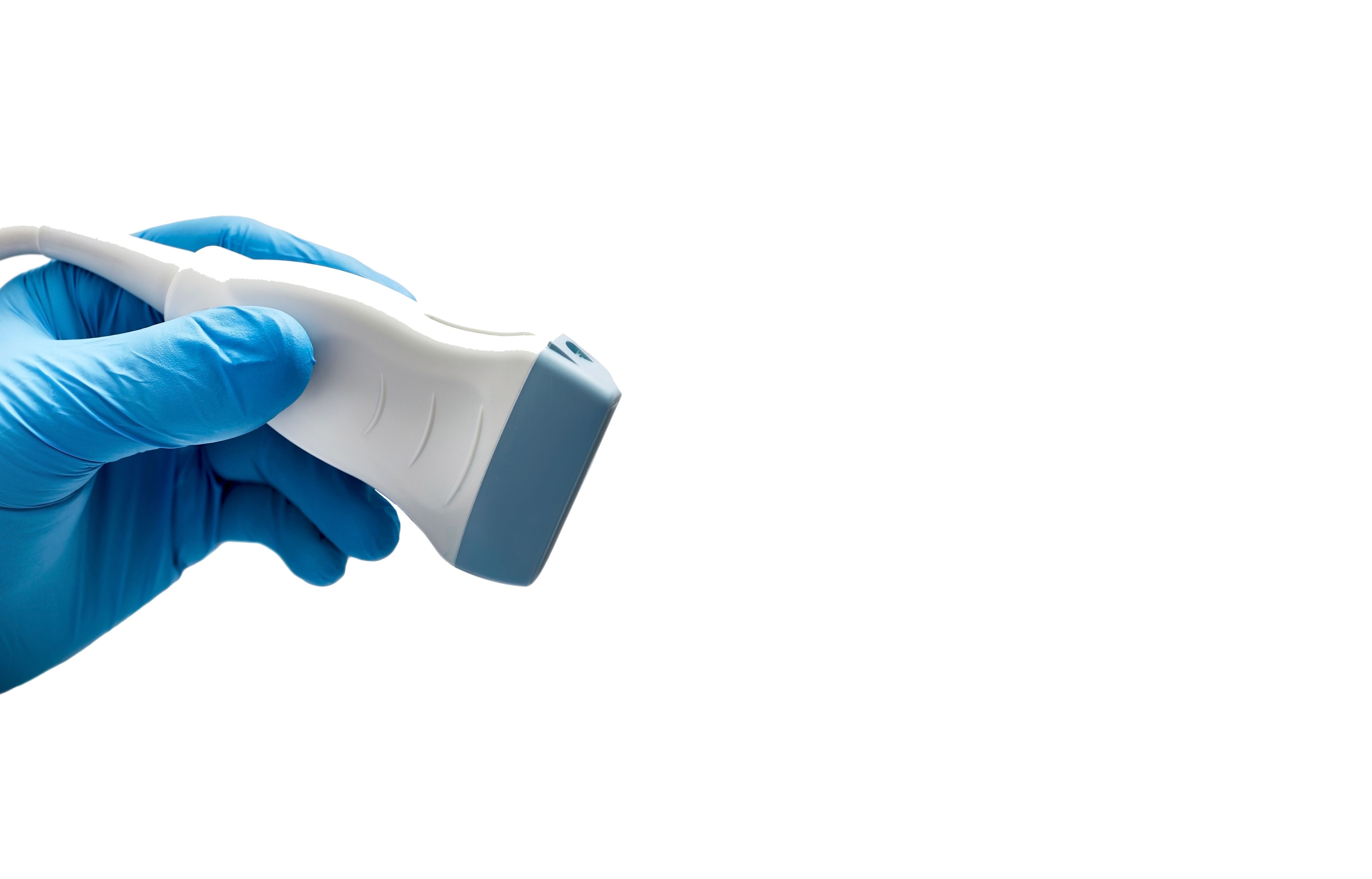
New ultrasound imaging to map drug delivery into the brain


Five years ago, ultrasound for drug delivery to the brain seemed like a black box to QBI’s Dr Pranesh Padmanabhan. But a conversation with collaborator Professor Jürgen Götz, who is developing a pioneering clinical trial program using non-invasive ultrasound to treat Alzheimer’s disease, sparked a question that would drive years of work: How does ultrasound actually work at a cellular level?

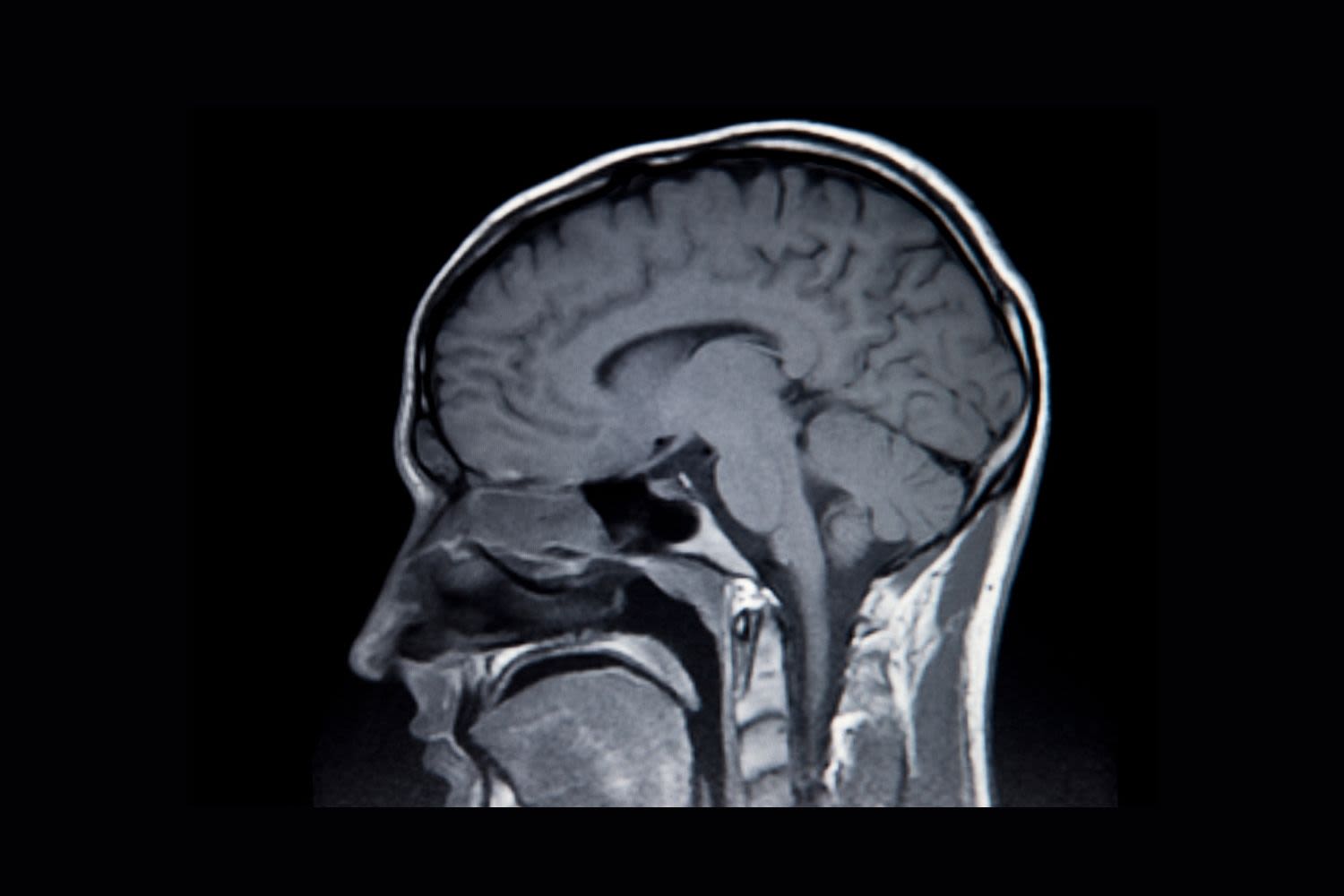
The goal was ambitious: find a safe way to open the blood-brain barrier – the protective shield that keeps most drugs out of the brain – using ultrasound. But to truly understand and refine the approach, Dr Padmanabhan's team needed to watch how cells responded to ultrasound in real-time.
"It seemed like an easy problem. Just put the device on the cells and see how they respond."

What started as a simple idea turned into five years of painstaking experiments. The team discovered that a brief 20-second burst of ultrasound could have effects lasting far longer than anyone expected – sometimes over an hour.
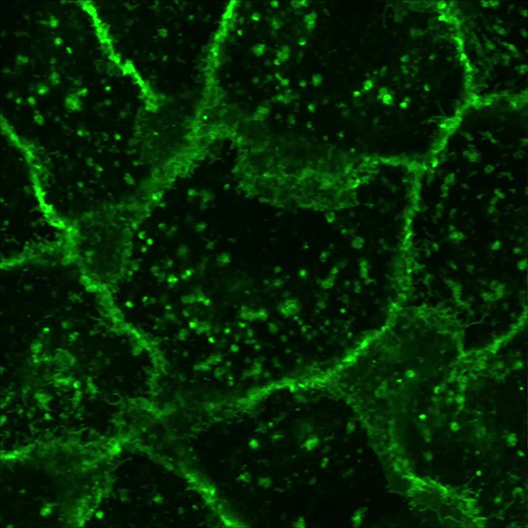
By pairing ultrasound pulses with microscopic ‘microbubbles’ attached to cell membranes, they could observe how bubbles oscillate, collapse and form tiny, temporary pores in cell surfaces. This process – known as sonoporation – lets drugs and other molecules slip into cells. But it can also damage cells if not properly controlled.
"One might think that a 20-microsecond treatment would only have a 20-microsecond effect, but that's not the case. We mapped how brief ultrasound can lead to long-lasting cellular changes."

First-author Mr Jonathan Lee, who led much of the live-cell imaging, highlighted the unexpected complexity behind these effects.
“What surprised us most was seeing how some cells recovered almost immediately while others showed delayed, subtle changes that weren’t obvious without continuous imaging. These hidden shifts might explain why some treatments cause side effects.”


Two cell fates: Life or death
Furthermore, their imaging revealed a split path for treated cells: some remained healthy, while others started shrinking and died within minutes. This ability to distinguish between these two outcomes opens up possibilities to fine-tune ultrasound doses.
“Now we can look at these two pathways. In cancer-targeted treatment, it’s fine to kill cells, but for blood-brain barrier opening, we need the opposite: we want cells to survive."

“We’re slowly moving towards quantitative, imaging-based pharmacology to not just find drug targets but understand exactly how drugs work at molecular and cellular levels.”
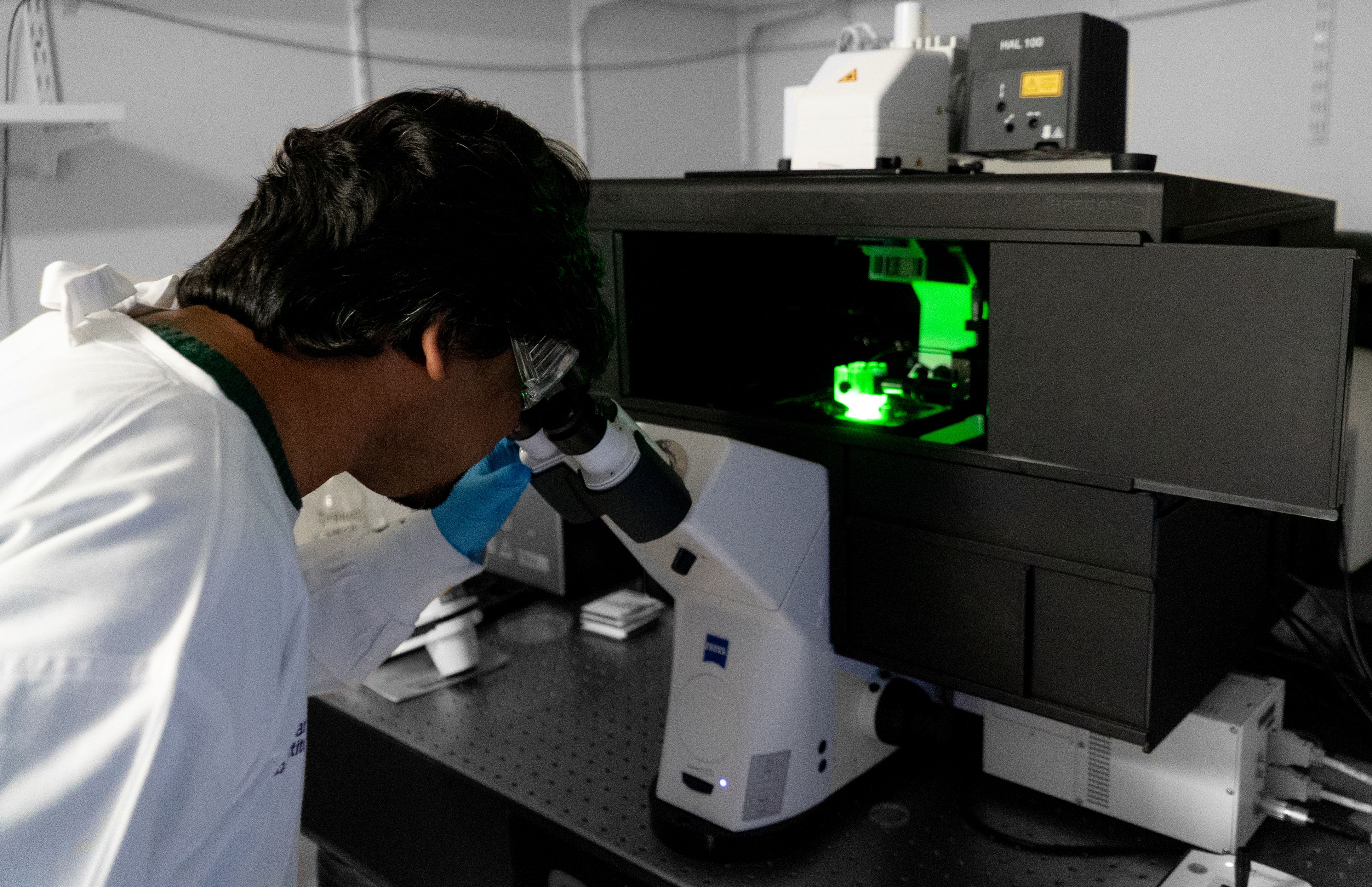
Live-cell imaging
QBI's Microscopy Facility Manager Dr Rumelo Amor detailed the challenges behind the scenes.
“Keeping the cells alive for long-term imaging while integrating the ultrasound device was the hardest part.”


Traditional live-cell setups need precise control of humidity, temperature, and CO2. The team had to modify these chambers to accommodate the custom-built ultrasound transducer. Fortunately, the LSM 980 NLO Airyscan 2 microscope at QBI had an extra-large incubation chamber that fit the custom setup. The Airyscan detector’s high-speed modes enabled the team to capture cellular events like vesicle movements and calcium spikes at both high temporal and spatial resolution.
“These fast-imaging multiplex modes were crucial because we needed to see rapid changes in cells, down to ~140 nanometres, in real time."


Collaborator in this research, QBI’s Clem Jones Centre for Ageing Dementia Research Director, Professor Jürgen Götz, highlighted the study’s broader significance.
“In using low-intensity ultrasound for drug delivery, the key is the underlying mechanism and whether drugs enter via an opened blood-brain barrier, vesicular transport or sonoporation. A crucial next step is determining whether sonoporation occurs in real brains treated with scanning ultrasound.”
He notes, this won’t be trivial but it’s vital for understanding and safely applying this emerging technology.

Mr Lee agreed, emphasising the importance of bridging lab models and patient treatments.
“We’ve built a powerful platform to dissect what happens at the single-cell level. The exciting challenge now is translating these insights to more complex, realistic brain environments.”


By revealing the hidden choreography of bubbles, pores and cellular responses, the QBI research team is laying the foundation for precise, safe and effective ultrasound-based therapies.
Their discoveries could one day turn ultrasound from a blunt tool into a scalpel of modern medicine, targeting disease with cellular precision.