Chapter 6
Small Brains
Research that helps us unravel the brain's inner workings will reveal what makes us human and pave the way for the development of treatments for some of the world's most debilitating brain conditions and disorders. In our growing and ageing society, age-related neurodegenerative conditions, such as dementia, pose an increasingly heavy social, economic and personal burden, highlighting the urgent need for brain research. These health conditions result from processes we must study in a living organism to advance our knowledge.
“What many people do not see is that behind every medical breakthrough that improves human health and wellbeing are the countless hours of research with animals. They give us remarkable insights into how the brain works and the potential ways we might fix it when it does not.”
Research on humans involves many ethical and technical obstacles. How do we study brain disease and mental disorders in humans? We are limited to minimally invasive techniques, including EEG, scans, or behavioural observations. We cannot sample the brain while someone is alive as we can for other organs.
In addition to being limited by how far we can interrogate the human brain, in research, it takes an exceptionally long time for results to materialise, not to mention the slippery ethical slope of conducting such experiments. Therefore, most of our researchers work with animals that allow more in-depth and time-efficient investigations than human subjects. So-called model organisms include rodents (mice and rats), fish, and even simple animals like flies and worms.
How can animals help us understand the human brain?
The answer to this question lies in the evolutionary origins of animal nervous systems, which date back several hundred million years to a time long before the divergence of animals into the likes of mammals, fish, insects, or worms. Thus, animal nervous systems today share many fundamental features and mechanisms with those of humans, and we can investigate them just as well, yet much more easily, than in a person.
Animals have shorter life cycles, meaning we can study them throughout their life or across several generations. Researchers can also control the animal's
environment (diet, temperature, exercise), allowing them to measure the impact of specific parameters. Researchers at UQ's Queensland Brain Institute adhere to strict standards and regulations for the humane, ethical and responsible treatment of animals.
“The molecules, the signalling pathways they use, and the basic biology around brain development and function are much the same. The basic rules we learn in flies and worms are directly applicable to humans. How the brain actually works in terms of cognition in humans is obviously harder to extrapolate from a fly, but for the wiring and how neurons work at that level, the fly, the worm, or the zebrafish are all good models.”

Rodents
Rodents like mice and rats make excellent model organisms due to their genetic similarities to humans and high reproductive rate. The mouse and the human genomes, for example, contain roughly 3.1 billion base pairs of DNA, of which only five per cent constitute protein-coding genes (required for making proteins—the building blocks of cells). While the similarity of these coding genes, at about 85 per cent, may seem low (compared to chimpanzees, with whom humans share about 98 per cent of the genome), the total repertoire of genes is virtually identical. This is one of the reasons why rodents are the most common
lab animals in fundamental and preclinical research.
At UQ's Queensland Brain Institute, researchers
use mice to test treatments and regimes before
trialling them on people. Examples include:
- Testing an experimental Alzheimer's treatment
based on ultrasound - Testing experimental drugs targeting a newly identified protein that causes motor neurone disease (MND)
- Studying the intensity and amount of exercise
required to improve cognition.

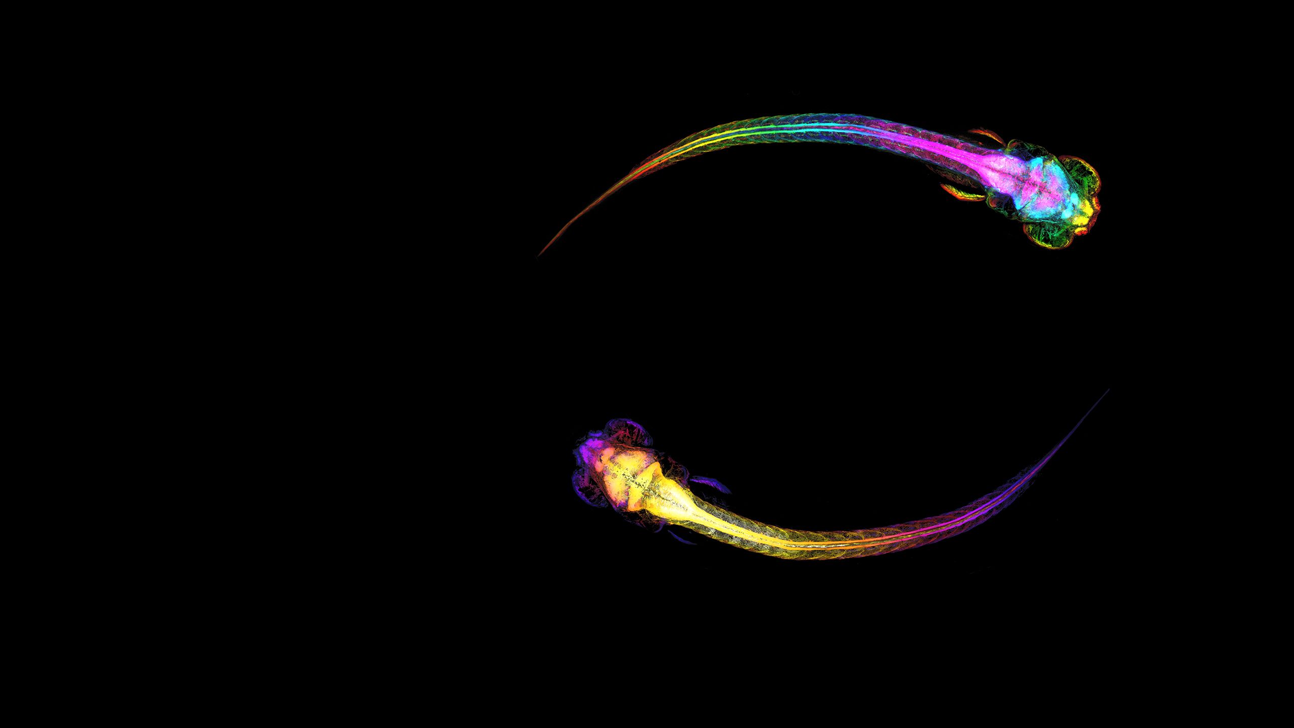
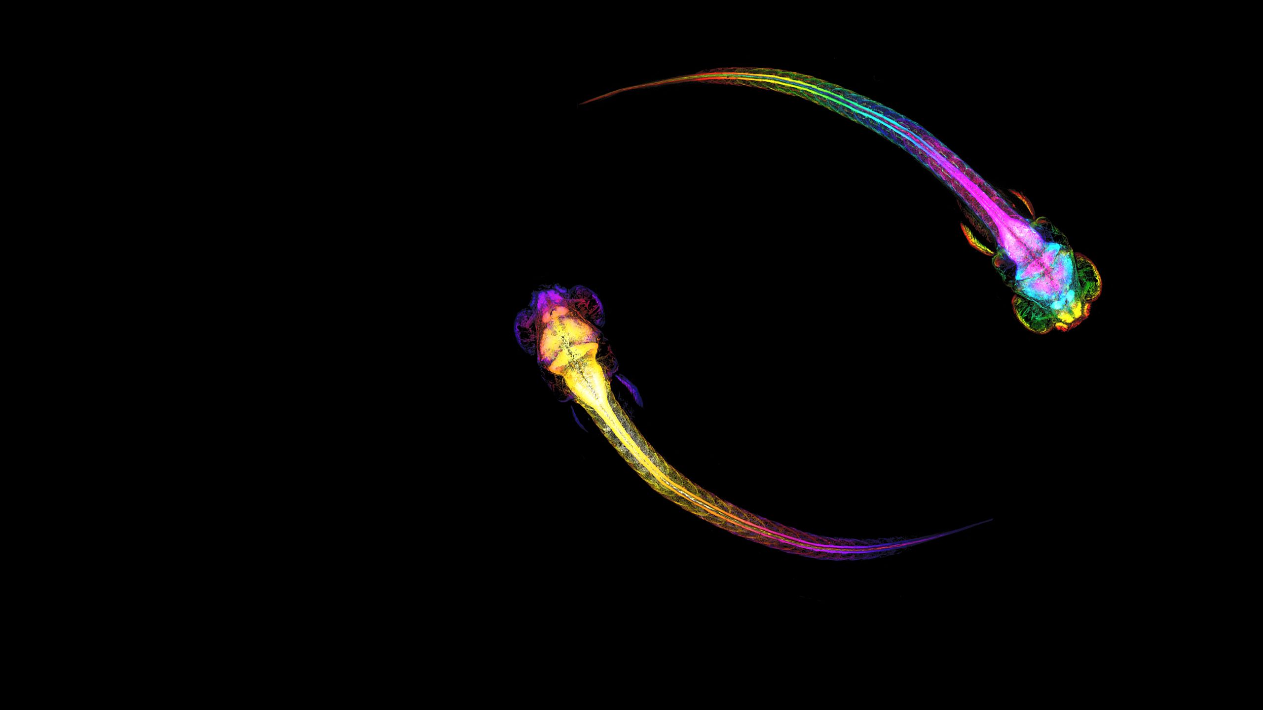
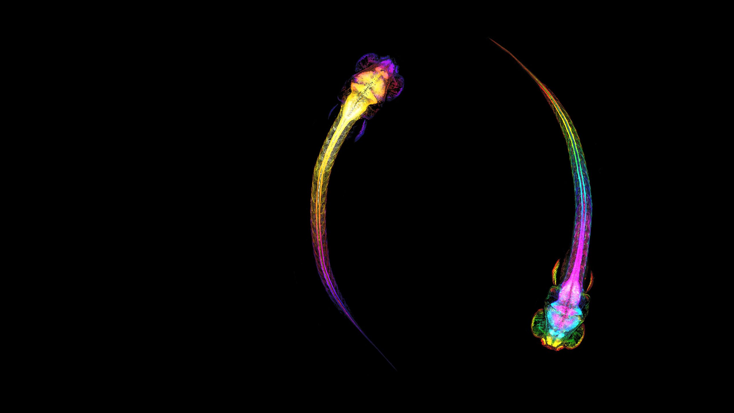
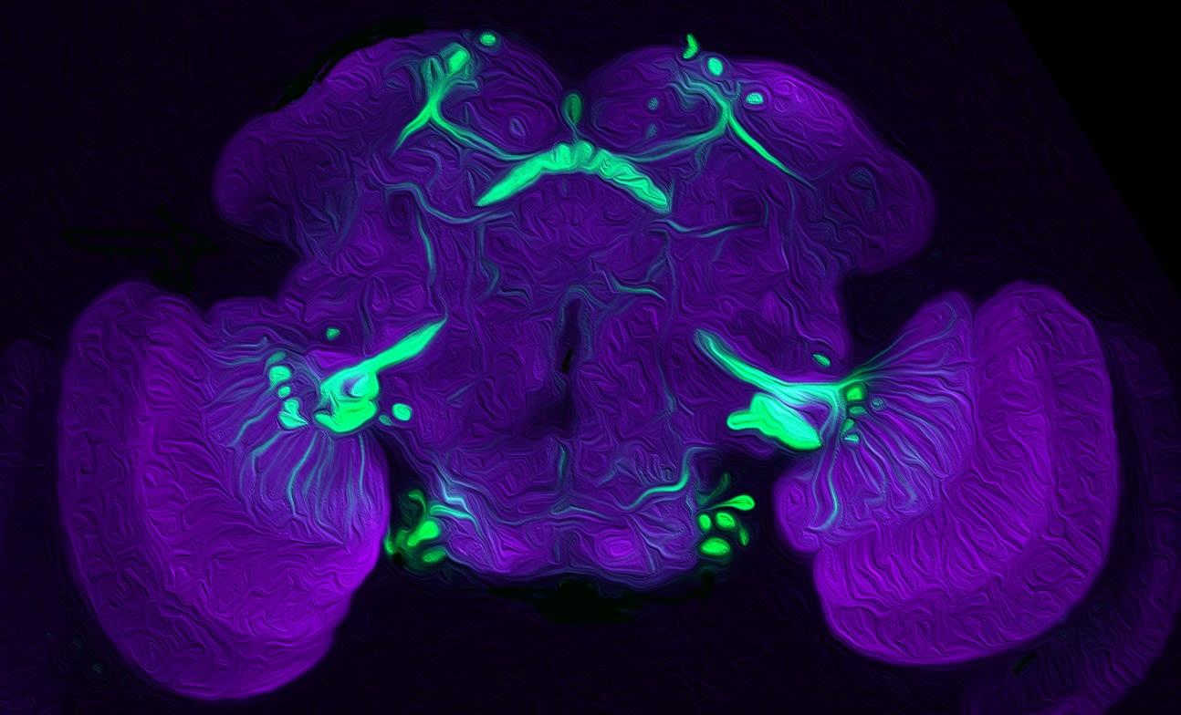
Fruit flies
The brains of insects, like that of the Drosophila or fruit fly, show remarkable structural and functional similarities to ours, albeit on a much smaller scale. The adult fruit fly brain contains only around 120,000 neurons compared to 100 billion in the adult human brain. Further, fruit flies exhibit complex behaviours despite their diminutive brain size.
Using the advanced genetic tools that the fruit fly system offers, researchers leverage these two features to understand the neuronal architecture and signalling within and between different brain areas and neuronal circuits.
The comparatively small number of fruit fly neurons is a huge advantage. Researchers can visualise individual neurons and their synaptic connections and record neuronal activity directly from the live fruit fly brain during different activities. This way, they seek to identify and understand highly conserved mechanisms governing brain circuitry and processing. Researchers at our Institute use these approaches to better understand what happens in our brains during different states of consciousness – wakefulness, sleep, and anaesthesia.
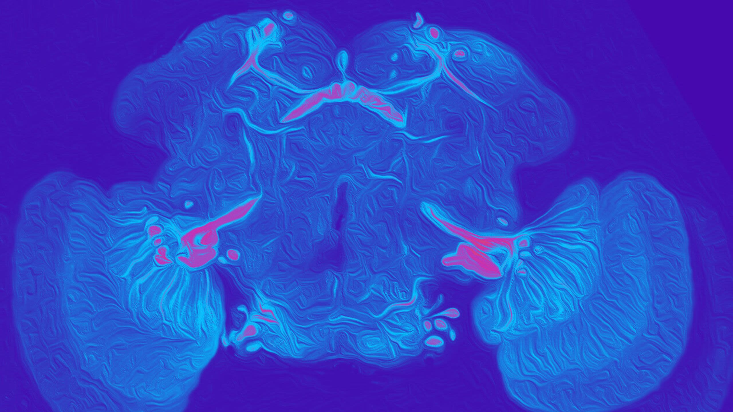

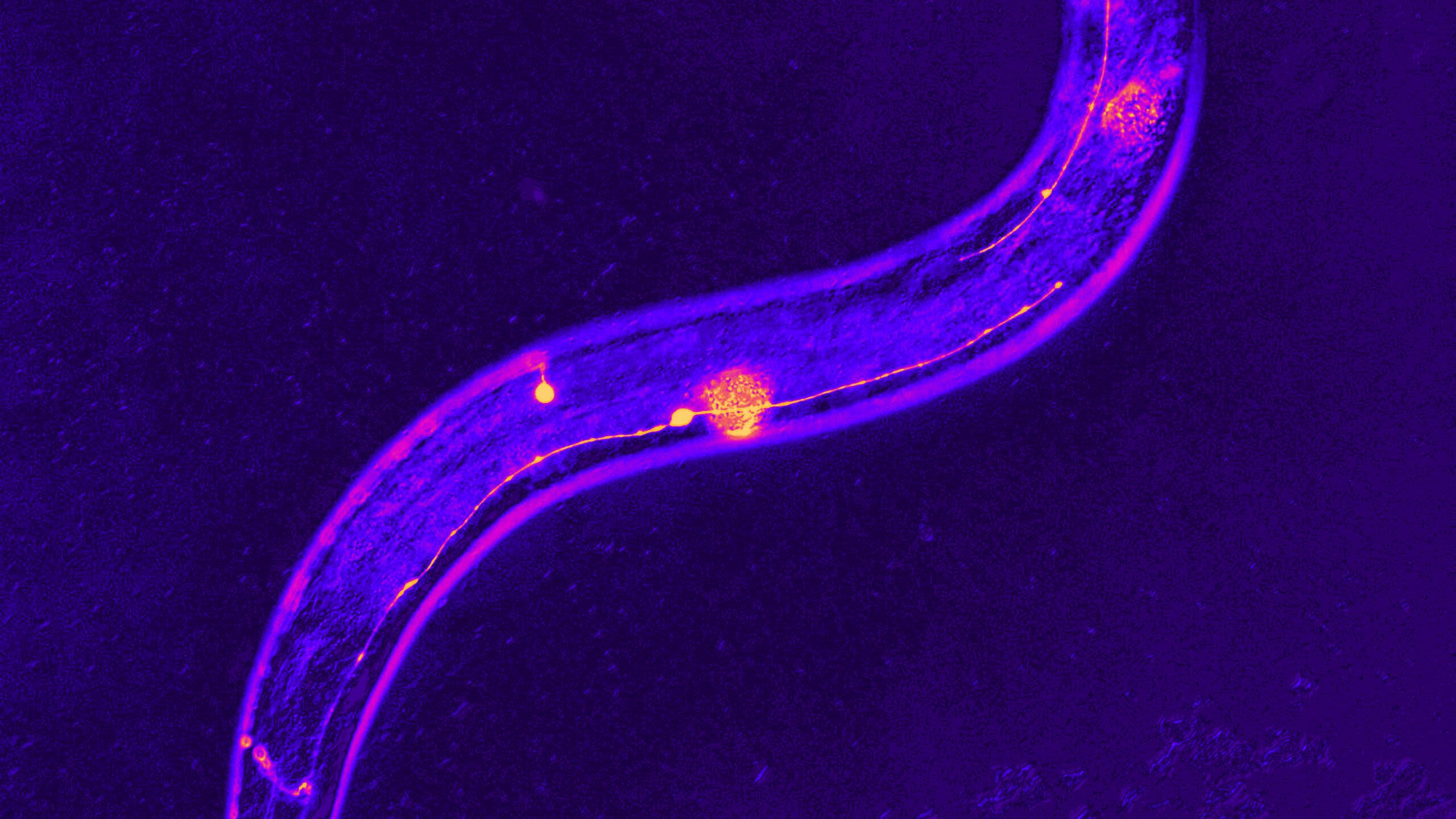
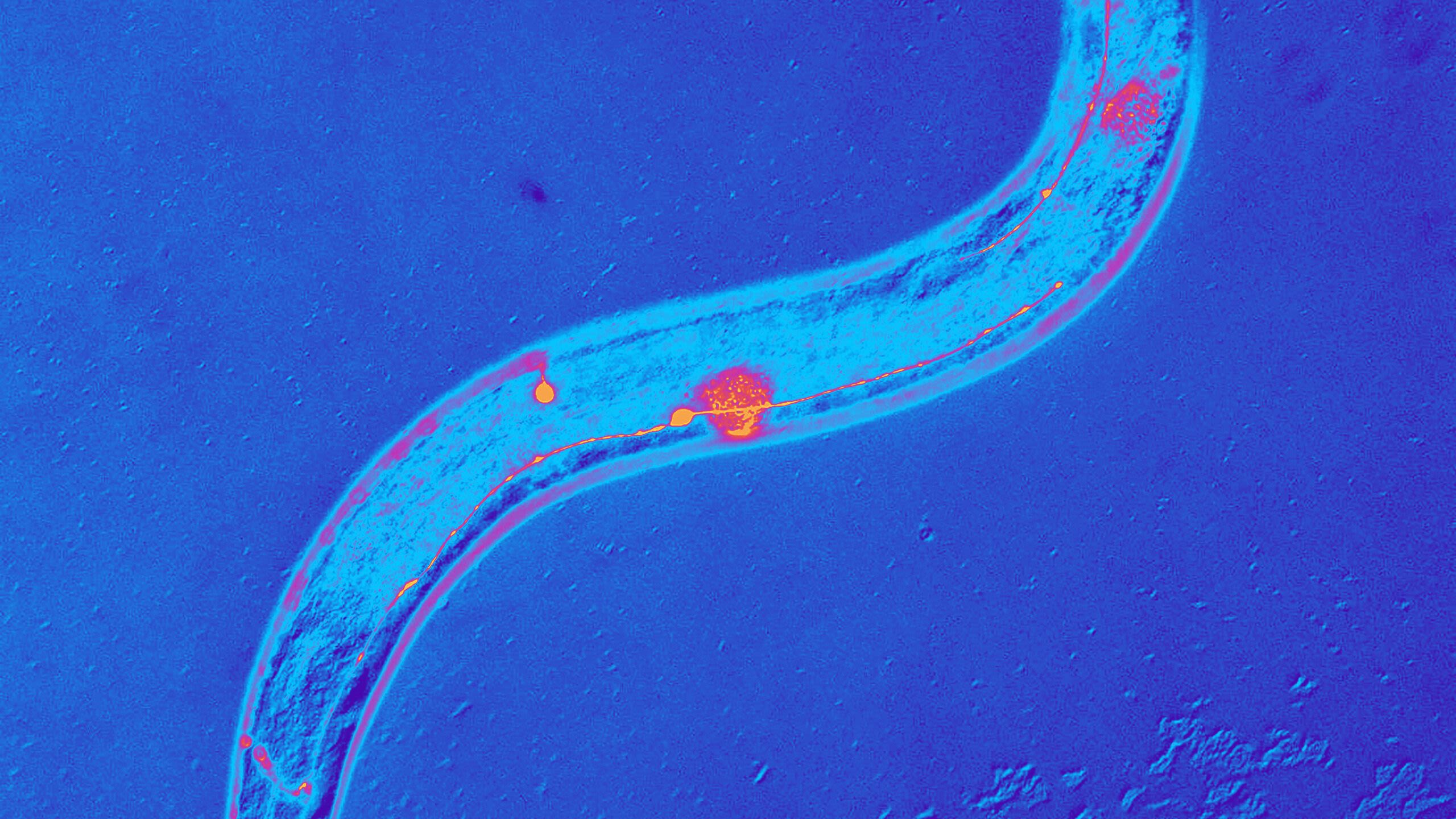
Worms
Caenorhabditis elegans is a nematode worm measuring a mere 1-2 mm long. Yet, it has a storied history as a classic model organism in biological and medical research. Many landmark discoveries, particularly on the development and function of the nervous system, were first made using this animal.
In 1998, it became the first multicellular organism with a completely sequenced genome and served as the proving grounds for the first-ever expression of green fluorescent protein (GFP)—a powerful tool to monitor gene expression—in a live organism.
The c. elegans body consists of only 959 cells, including 302 neurons, which can be readily observed due to their transparent body. Its genome shares around 60-80% of its protein-coding genes with humans. C. elegans has a lifespan of 12 to 18 days, which allows scientists to generate large sample sizes and results in a fast turnaround for experiments. Researchers can easily implement and observe the effects of genetic modifications likely to involve similar molecules and mechanisms as they would in humans.
At UQ's Queensland Brain Institute, researchers use c. elegans to:
- Better understand the cellular and molecular processes of learning and memory
- Study age-related conditions associated with mitochondrial dysfunction (mitochondria produce the energy used by all cells)
- Explore how viruses impact the brain
- Investigate axonal repair mechanisms that could lead to treatments for conditions arising from nerve damage, such as glaucoma or paralysis.