Chapter 5
Technology Turbocharging Research
The brain is an organ encased in the skull and composed of vast numbers of cells and connections. Interrogating the brain's structure and function is fundamentally challenging, and much of the growth in neuroscience has been driven by developments in technology.
1. Next-generation sequencing
Since its commercialisation in the early 2000s, next-generation sequencing (NGS) technology has revolutionised neuroscientists investigate the fundamental molecular mechanisms underlying brain function.
This process has revealed many previously unknown disease-causing genes, laying the groundwork for the development of highly specific therapeutics. Previous sequencing technologies were either limited in maximum sequence length or very time-consuming. NGS overcomes both of these problems.
2. Gene editing (CRISPR/Cas9)
Gene editing has been widely used in fundamental and clinical research and holds enormous potential for the novel therapeutic approaches for many inherited diseases. Only recently, however, has the term caught the public eye. This is mainly due to the rise of CRISPR, a Nobel Prize-winning (2020) gene-editing technology, derived from the viral defence system of bacteria.
The CRISPR system spectacularly outperforms older gene editing platforms as it is faster, cheaper, easier to use, and vastly more efficient. Where older methods required researchers to make new primers for each new target sequence — a very lengthy and expensive process — CRISPR can identify its target sequence via so-called guide RNAs, short nucleic acid sequences that are easy and cheap to produce. Because of its low cost, ease of use, and unprecedented editing efficiency, scientists laud CRISPR technology as the trailblazer in the gene-editing age.


3. Advanced Imaging Technologies
Evolving imaging technologies provide neuroscientists increasingly more detailed insight into the brain's neuronal architecture, synaptic connections and how they are made and work.

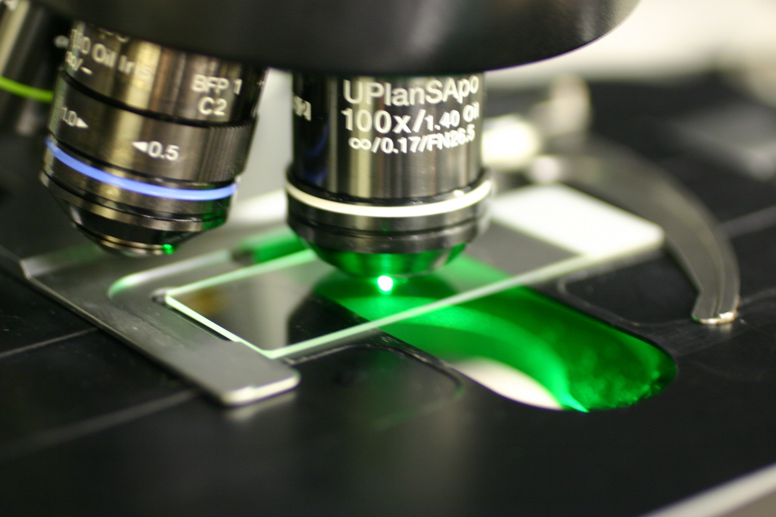
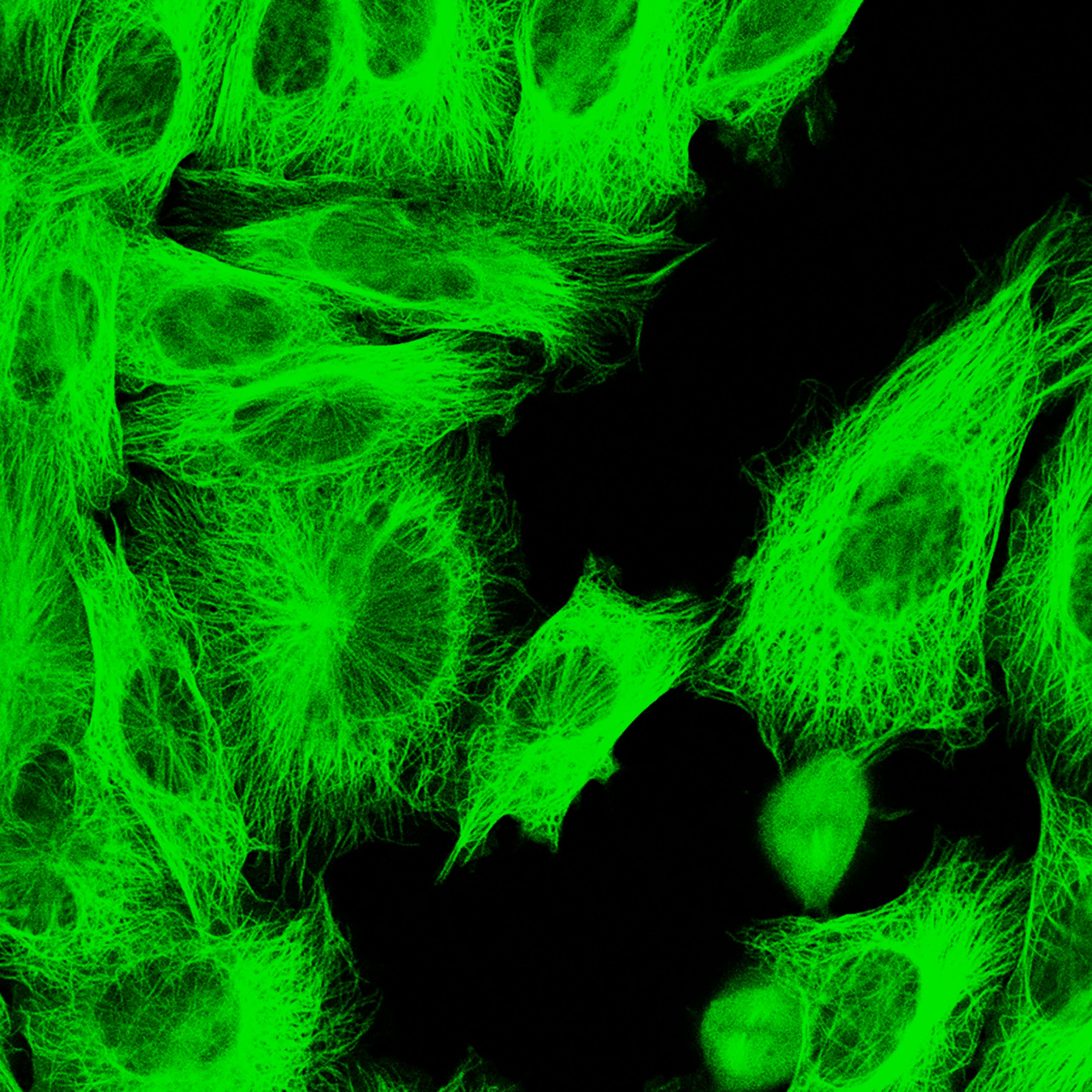
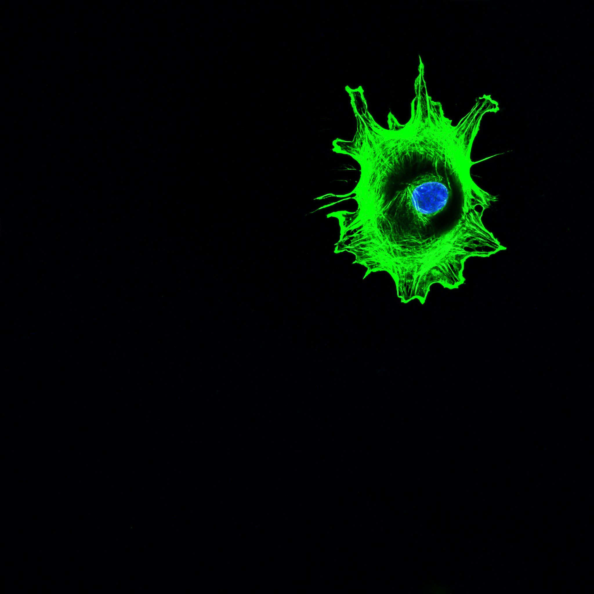
1. For decades, fluorescence microscopy, with its versatility in labelling and imaging different cell types, molecules, and even gene expression has been a cog in the wheel of biological and biomedical research. Unfortunately, its optical capability that allows fluorescent dyes to be used in the first place also limits its resolution to the sub-micrometre scale, meaning that many smaller proteins, which play critical roles in neural function, were beyond the resolving power of fluorescent microscopes. That is until the invention of Super Resolution Microscopy (SRM), a Nobel Prize-winning (2014) optical imaging method that shattered the limits of the physically possible. With SRM, researchers can now perform live-cell and three-dimensional imaging at nanometre resolutions, a realm previously reserved for electron microscopes.
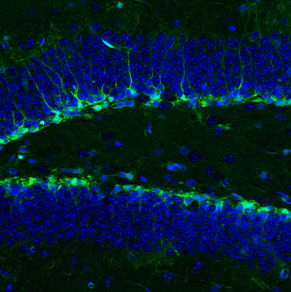
2. First patented in 1990, two photon microscopy, another advancement on 'traditional' fluorescence microscopy, dramatically reduced background signal since its beam of excitation light is much more focused on the viewed plane. Because of this, researchers can observe cells and molecules up to one millimetre deep in live tissue and even in entire organisms, such as the fruit fly. This makes it possible to track particle movement inside or between cells during specific behaviours and helps us understand cellular interactions and neural signalling.
“This technology is invaluable in understanding receptor dynamics on the surface of neurons and helping reveal the mechanisms underlying synaptic connectivity and memory acquisition.”
3. Single Particle Tracking (SPT) is a powerful tool that allows neuroscientists to observe the movements of single fluorescently labelled molecules as they move in or around cells. This can, for example, be invaluable in understanding receptor dynamics on the surface of neurons and help reveal mechanisms underlying synaptic connectivity and neuronal signalling. ither scanned into or drawn directly into a computer system.
4. Functional magnetic resonance imaging (fMRI) first developed in the 1990s, has proven invaluable in our efforts to understand which brain regions serve which cognitive functions. Like ordinary MRI, fMRI creates high-resolution images of soft structures in our bodies, such as the brain. However, fMRI also measures the small changes in blood flow that occur with brain activity. Neuroscientists can use this information to link regional brain activity with concurrent cognitive tasks. On the other hand, clinicians use fMRI to evaluate the effects of stroke, concussion, or other conditions that may have affected a patient's brain function.


4. Machine learning/AI tools
Given the immense amounts of data obtained from advanced research methods, such as those listed above, the emergence of machine learning and artificial intelligence (AI) tools has been invaluable and timely. AI is transforming neuroscience research by enabling efficient analysis of complex brain data, such as highly detailed images and scans or complex brain activity signals.
As such, machine learning algorithms and AI tools are immensely useful for tasks such as brain mapping, disease diagnosis, drug discovery, and treatment optimisation, and the development of brain computer interfaces.

Dr Clarissa Whitmire
“There's been a massive explosion of experimental tools in neuroscience. We've gone from recording the activity of individual neurons, which transformed our view of neuroscience in the last 50 years, to now recording hundreds or thousands of neurons simultaneously. The ability to not only record, but precisely control, populations of neurons will lead to transformational shifts in our understanding of the brain.”
In 2023, neuroengineer* Dr Clarissa Whitmire joined UQ's Queensland Brain Institute as a senior research fellow and is the Institute's newest group leader. In a joint appointment with UQ's School of Engineering, Clarissa seeks to combine her expertise in biomedical engineering with innovative neuroscience to decipher how the brain encodes and processes information about the external world in distributed populations of neurons.
The time to tackle ambitious problems has arrived, Clarissa says, thanks to staggering advances in in recording, imaging, and genetic methods.
Originally from the United States, Clarissa arrived at UQ following a postdoctoral appointment at the Max Dellbruck Center for Molecular Medicine in Berlin. She obtained her bachelor's in science (Biomedical Engineering) from North Carolina State University and PhD in Biomedical Engineering in a joint program between the Georgia Institute of Technology and Emory University in 2017.
In her research, Clarissa combines innovative electrophysiology, imaging, and genetic tools with computational approaches to develop models of information representation, processing, and transmission within and between neural circuits.
The internal representation of the outside world is built on patterns of neural activity, commonly referred to as the ‘neural code’. While we often model the neural code as a linear mapping from stimulus to spikes, it is extremely complicated and nonlinear, even very early in the sensory pathway.
In particular, the neural code explodes in complexity at the thalamocortical circuit, where nearly all sensory information travels through the thalamus before reaching the cortex. These highly interconnected structures form a functional unit that underlies our ability to perceive. Dysfunction in the thalamocortical circuit has been implicated in several disorders, including schizophrenia and autism spectrum disorder.
The challenge, she explains, lies in making sense of the immense amounts of data that these new tools allow researchers to collect—a challenge she intends to tackle by applying engineering frameworks to the biological circuits formed by the neurons in our brains.
*Neuroengineering involves using and developing engineering techniques to understand, interact with, or influence neural function.