Solving big-data bottlenecks for microscopy


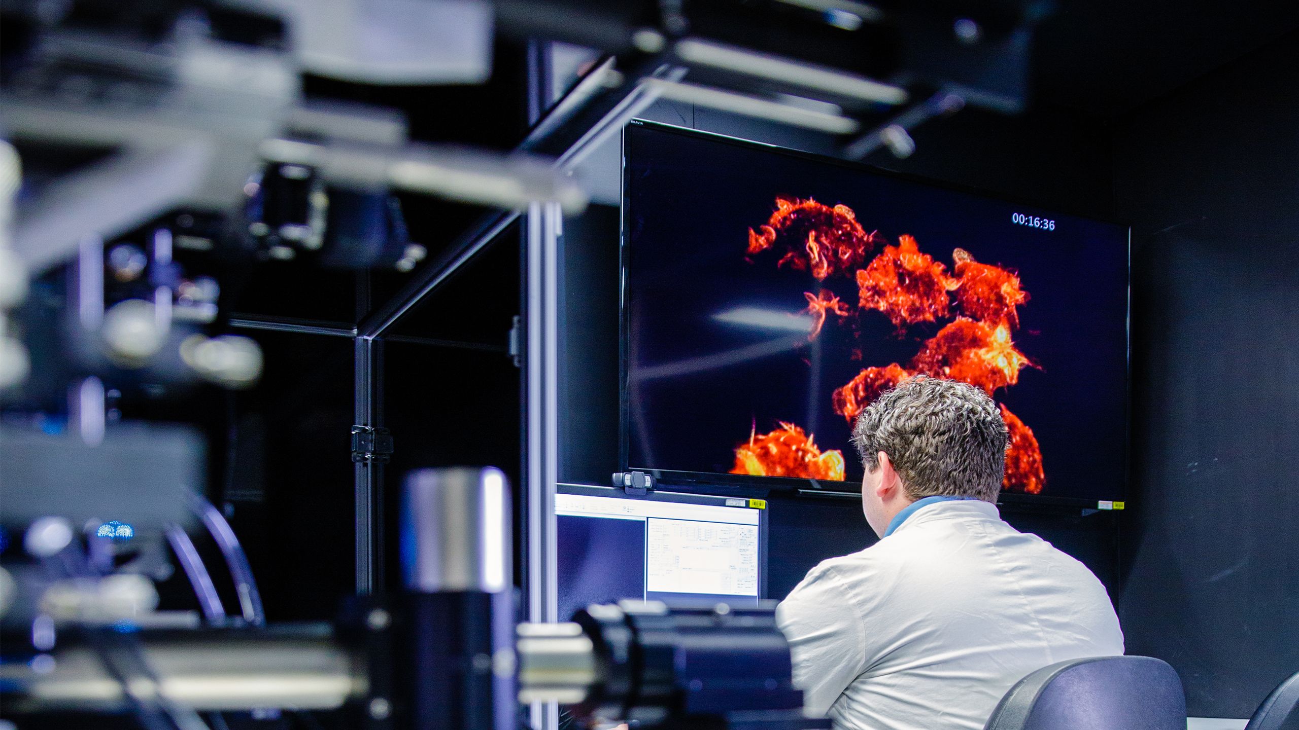
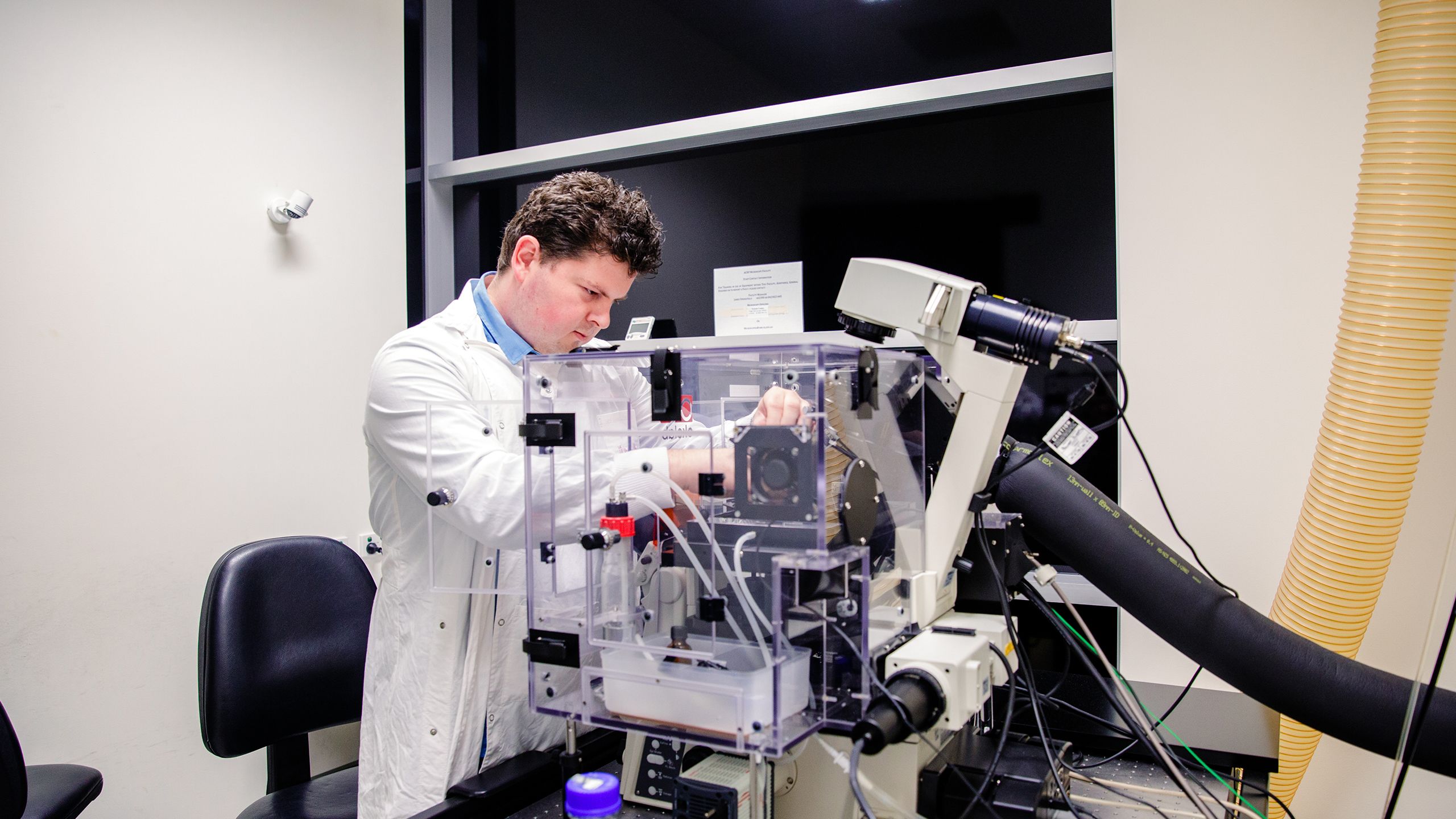
Nicholas Condon working in the ACRF:Cancer Biology Imaging Facility, using the 3i Lattice Light-Sheet microscope to capture high-speed volumes of macrophages sampling their environment. This microscope can produce many terabytes of RAW data per day requiring extensive post acquisition analysis (Image: IMB Microscopy).
Nicholas Condon working in the ACRF:Cancer Biology Imaging Facility, using the 3i Lattice Light-Sheet microscope to capture high-speed volumes of macrophages sampling their environment. This microscope can produce many terabytes of RAW data per day requiring extensive post acquisition analysis (Image: IMB Microscopy).


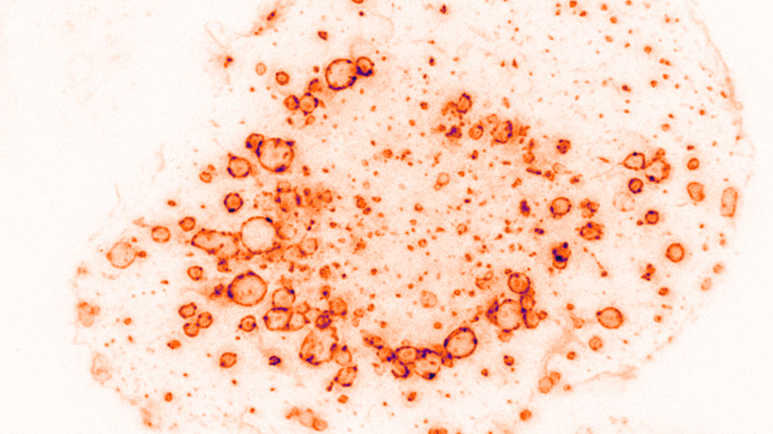
Immune cells continuously sample their environment, taking up gulps of the extracellular space into structures called endosomes. In this example the many endosomes can be observed with the help of a lipid specific label (Image: Nick Hamilton).
Immune cells continuously sample their environment, taking up gulps of the extracellular space into structures called endosomes. In this example the many endosomes can be observed with the help of a lipid specific label (Image: Nick Hamilton).


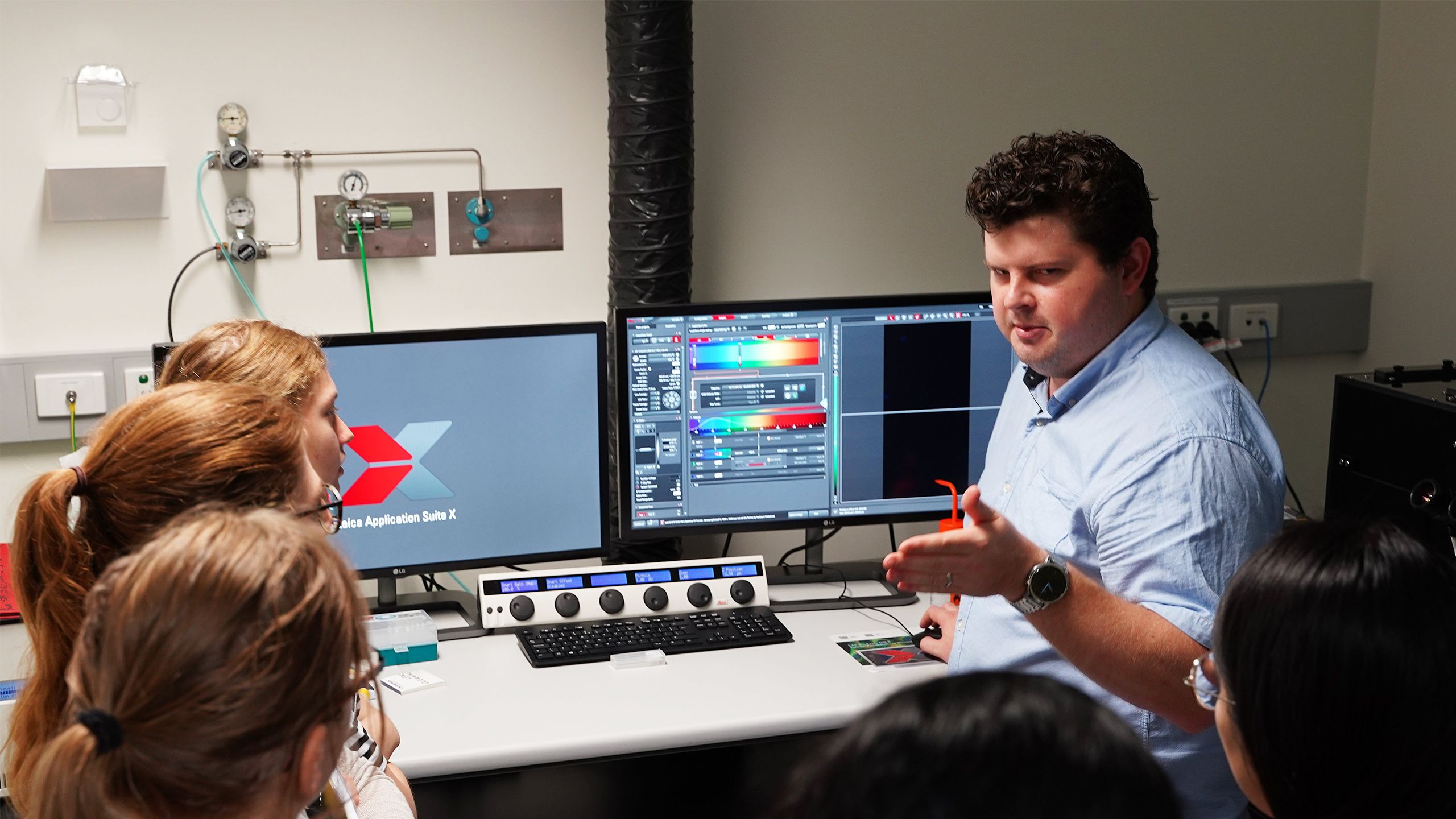
Nicholas Condon teaching meeting attendees how to acquire a confocal image using the Leica SP8 Confocal microscope as part of the Light Microscopy Australia meeting in Brisbane 2019. Photo Credit: Nick Hamilton (Image: IMB Microscopy).
Nicholas Condon teaching meeting attendees how to acquire a confocal image using the Leica SP8 Confocal microscope as part of the Light Microscopy Australia meeting in Brisbane 2019. Photo Credit: Nick Hamilton (Image: IMB Microscopy).
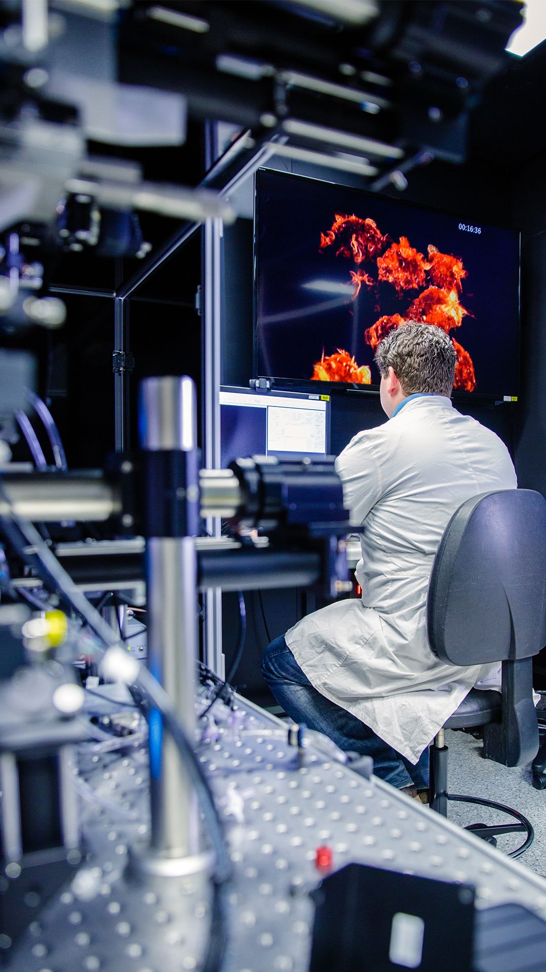
Nicholas Condon working in the ACRF:Cancer Biology Imaging Facility, using the 3i Lattice Light-Sheet microscope to capture high-speed volumes of macrophages sampling their environment. This microscope can produce many terabytes of RAW data per day requiring extensive post acquisition analysis (Image: IMB Microscopy).
A University of Queensland imaging scientist has secured more than $800,000 (US$595,000) to help unlock scientific discoveries by improving the use of big data from Australia’s most advanced microscopes.
UQ Institute for Molecular Bioscience’s Dr Nicholas Condon has received a five-year grant from the Chan Zuckerberg Initiative, which supports imaging experts who help to better understand what causes disease and how to develop treatments.
“With recent advances in light microscopy, we are now capturing larger and larger volumes of data than ever before,” Dr Condon said.
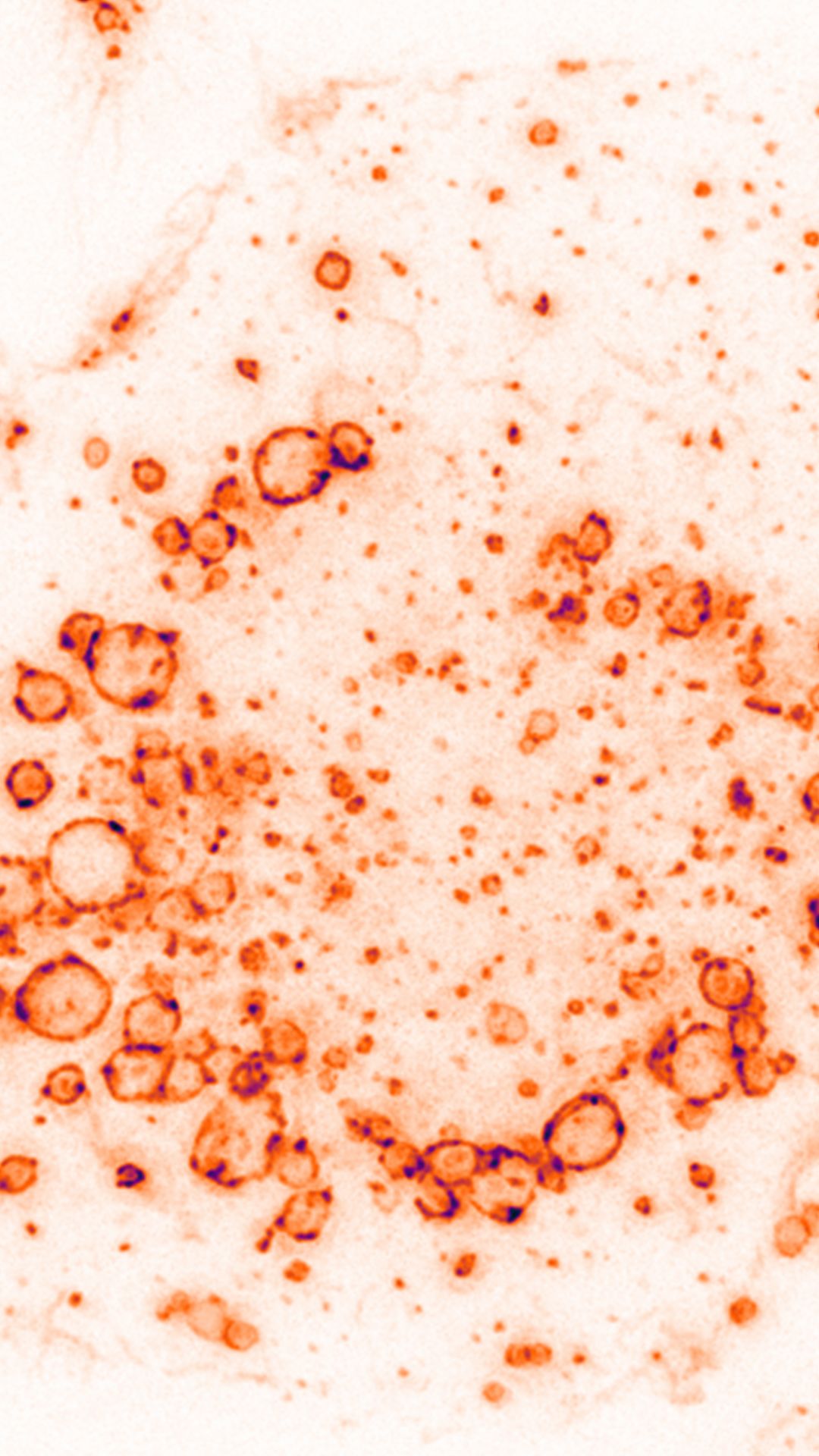
Immune cells continuously sample their environment, taking up gulps of the extracellular space into structures called endosomes. In this example the many endosomes can be observed with the help of a lipid specific label (Image: Nick Hamilton).
“We can now watch a cell over a period of time, for example many hours, seeing how it moves and responds to different stimuli, and this creates terabytes of data.
“My aim is to speed up and automate processing of microscopy data, using an ‘image processing portal’—we are developing tools that speed up mundane tasks, freeing up time for making discoveries.”
The Chan Zuckerberg Initiative (CZI) recently awarded $43 million (US$32 million) to support biomedical imaging advances, including technology development.

Nicholas Condon teaching meeting attendees how to acquire a confocal image using the Leica SP8 Confocal microscope as part of the Light Microscopy Australia meeting in Brisbane 2019. Photo Credit: Nick Hamilton (Image: IMB Microscopy).
Dr Condon will assist researchers from many disciplines including biology, chemistry, physics, engineering and ecology to use advanced microscopic methods to answer questions such as how immune cells respond to bacteria and how drugs can be delivered into cells, and to create tools to study human pathogens.
“Our vision is for the IMB Microscopy Facility to be the leading biological imaging and image analysis centre in Australia to advance the science and scope of imaging and microscopy," he said.
“This award will really help us advance towards this goal and improve the technology and techniques available to Australian scientists.”
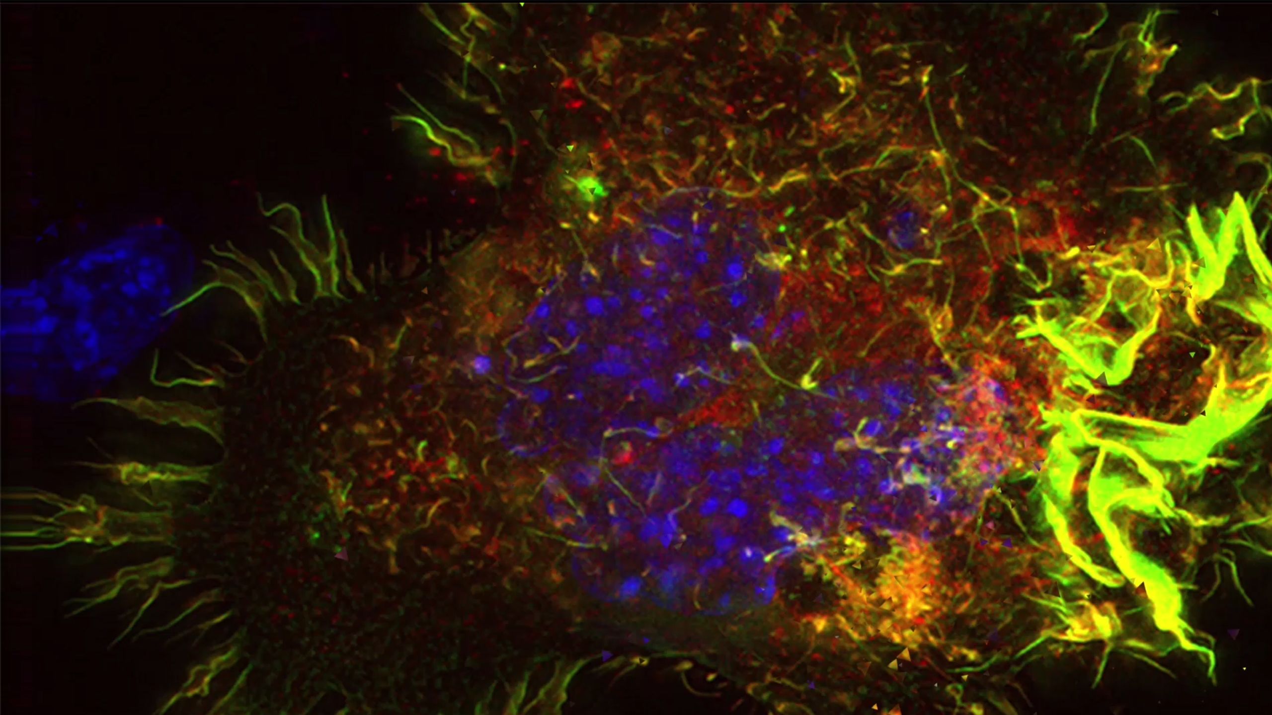
Lattice Light-sheet microscopy allows for rapid high-speed volumetric imaging not possible on other systems. In this example, an immune cell can be observed engulfing a foreign object in a process called phagocytosis in exquisite detail (Video: Nicholas Condon & Nyakouy Yak).
In addition to helping other researchers, Dr Condon uses advanced imaging methods, such as the lattice light-sheet microscope, to pursue his interest in the dynamic membrane environment of immune cells.
He identified a new mechanism for macropinocytosis — how cells drink and eat — which may have implications for manipulating the immune system in disease.
IMB Director Professor Ian Henderson said Dr Condon had successfully established lattice light-sheet microscopy at IMB and spearheaded developments in hardware, software and training.
“This initiative conceived by Dr Condon will empower microscopy users to take control of their large datasets, speeding up handling and analysis of big data, and ultimately accelerate the scientific process,” Professor Henderson said.
Founded by Dr Priscilla Chan and Mark Zuckerberg in 2015, the Chan Zuckerberg Initiative leverages technology to help solve some of the world’s toughest challenges — from eradicating disease to improving education and reforming the criminal justice system.


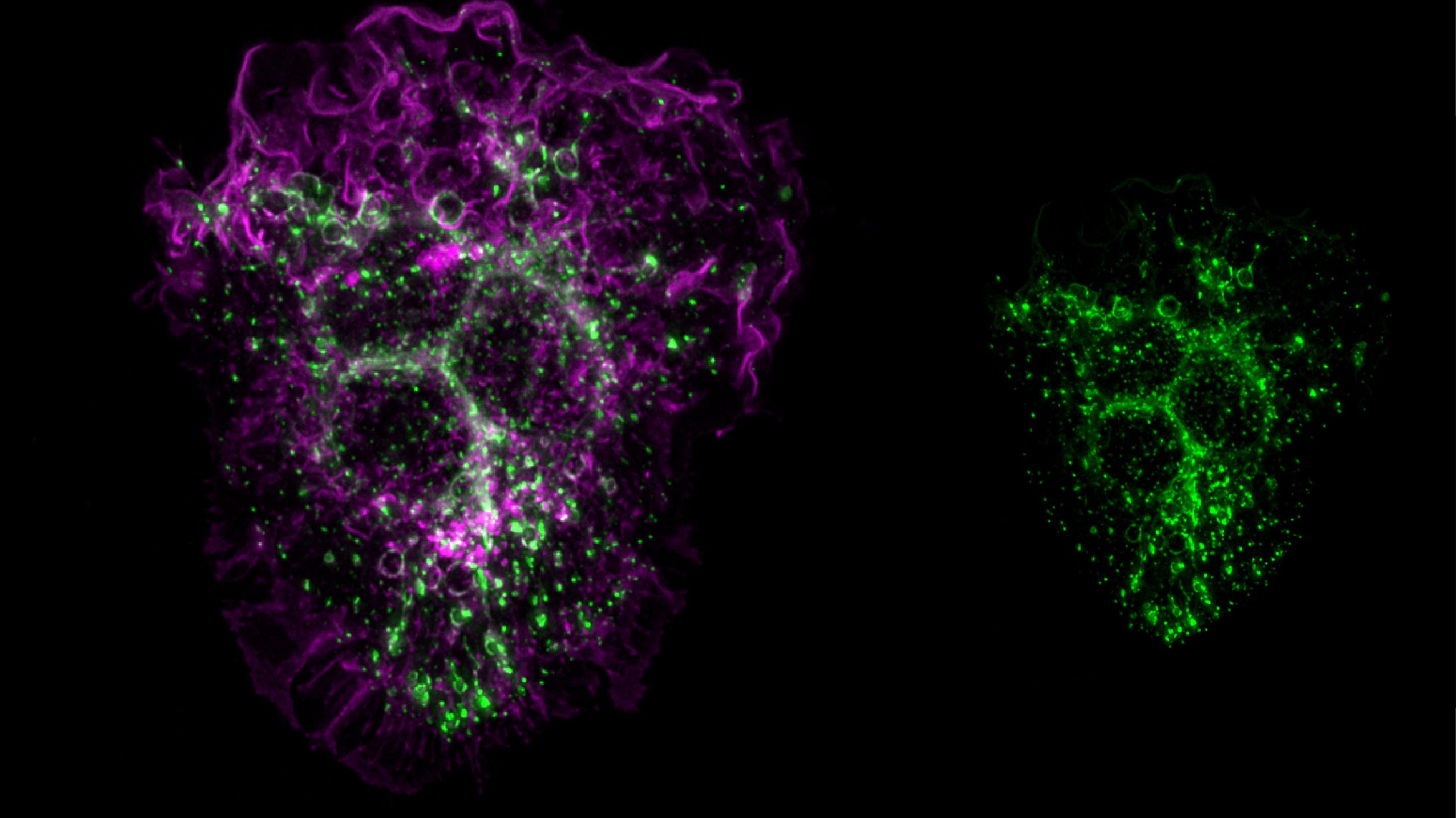


Mouse macrophage showing a large dorsal ruffle structure used to sample its environment to detect and ingest foreign objects such as pathogenic bacteria (Image: Nicholas Condon).
Mouse macrophage showing a large dorsal ruffle structure used to sample its environment to detect and ingest foreign objects such as pathogenic bacteria (Image: Nicholas Condon).


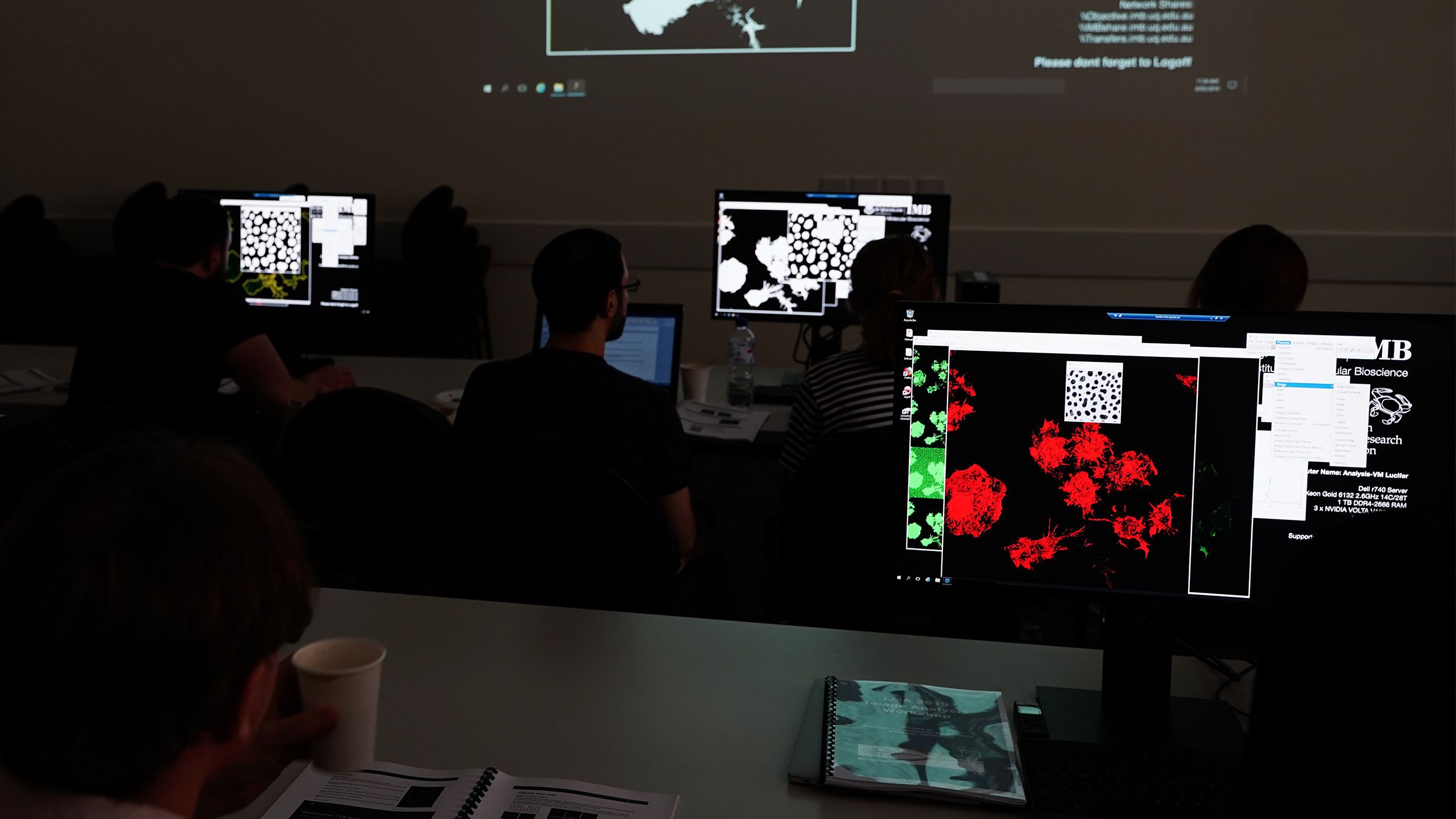
Image analysis workshop presented as part of the Light Microscopy Australia society meeting in Brisbane 2019 (Image: Nick Hamilton).
Image analysis workshop presented as part of the Light Microscopy Australia society meeting in Brisbane 2019 (Image: Nick Hamilton).


Nicholas Condon working in the ACRF:Cancer Biology Imaging Facility (Image: IMB Microscopy).
Nicholas Condon working in the ACRF:Cancer Biology Imaging Facility (Image: IMB Microscopy).
Media:
Dr Nicholas Condon, n.condon@uq.edu.au, +61 (0)4 0090 9510; IMB Communications, communications@imb.uq.edu.au, +61 (0)4 0566 1856