Growing neurons
The BRAIN magazine
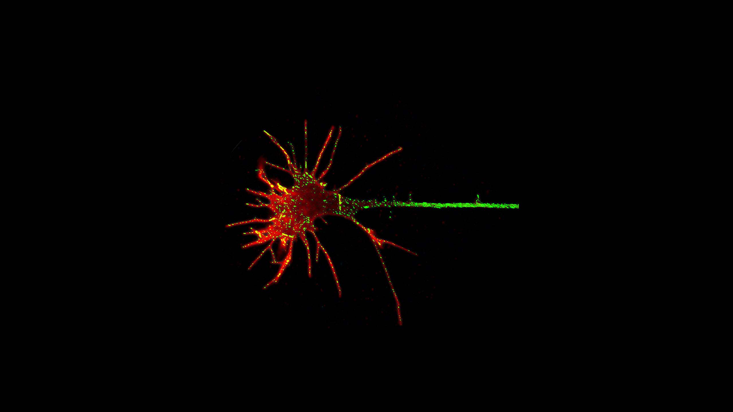

In the developing brain, neurons are born and then migrate to their final destination in the brain. Once at their destination, they extend out the long cable called the axon to start forming connections with other neurons.
The direction of axon migration is determined by the response of the growth cone at the tip of the growing axon to chemical cues in the local environment. These cues guide the direction of the growing axon by either attraction or repulsion.
Brains in a dish
For researchers to understand how the brain works and how it malfunctions in diseases and injuries, they study the brain in a setting as close as possible to its natural environment. To some extent, researchers can do this by taking slices of brain tissue to briefly study the biology of neurons while the cells are alive.
Neurons and glial cells can also be separated and studied individually in a laboratory dish. Recently, researchers like Professor Ernst Wolvetang from UQ’s Australian Institute for Bioengineering and Nanotechnology have been creating a more realistic three-dimensional environment using mini-brain-like organs called brain organoids. These are miniature models with similar cellular make-up and architecture as a developing human brain, mimicking many aspects of early development and enabling researchers to discover the multiple cellular mechanisms involved in brain formation.

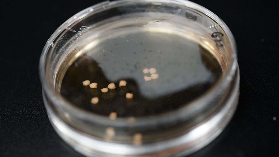
Organoids in a Petri dish. Image: Australian Institute for Bioengineering and Nanotechnology
Organoids in a Petri dish. Image: Australian Institute for Bioengineering and Nanotechnology
Organoids are made by taking adult human cells from the skin, hair follicles or blood and culturing them in a special growth medium to create stem cells which, after exposure to the correct growth factor, generate neural stem cells. These cells can then be triggered to produce self-organising structures called organoids, which contain many populations of neurons and exhibit a basic architecture similar to that seen in a developing brain.
Studying organoids will allow researchers to understand the progressive molecular and cellular changes that may occur in several developmental disorders and potentially screen existing, new and emerging medicines for efficacy and possible side effects. Organoids are also now being used to study neurodegenerative diseases such as Alzheimer’s disease.


Organoids under a microscope. Image: Australian Institute for Bioengineering and Nanotechnology
Organoids under a microscope. Image: Australian Institute for Bioengineering and Nanotechnology
Definitions
Differentiation
The process whereby a stem cell develops into a specific cell type.
Axon
The long, thin, cable-like structure of a neuron. Electrical impulses are sent along the axon to the dendrites of a nearby cell.
Growth cone
This highly sensitive, dynamic and pathfinding tip is essential for growing neurons and rewiring circuits after nerve cell damage.
Dendrite
These small branch-like projections of a neuron receive information sent via the axon.
Synapse
The junction between the axon of one neuron and the dendrite of another, through which the two neurons communicate.